Case Study Image

AI-powered brain tumor segmentation for surgical planning

Client:

Mivora Neuroscience Institute (USA)

Type:

Specialized Neurology

Annual imaging volume:

120,000+ brain MRI scans

Challenge:

Time-consuming manual tumor segmentation, variability in surgical planning

Mivora Neuro Hospital, a renowned neurosurgical institute in Switzerland, faced growing challenges in accurately segmenting brain tumors during pre-surgical planning. Manual segmentation performed by radiologists required extensive time—often several hours per case—and carried the risk of variability in identifying tumor boundaries. These inconsistencies could impact surgical precision and overall patient outcomes.

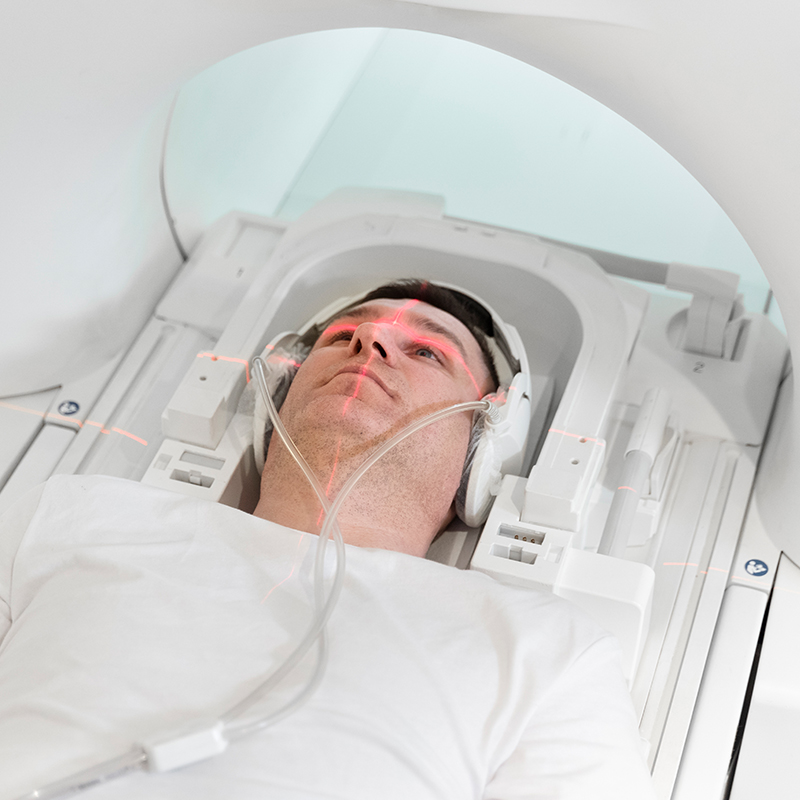
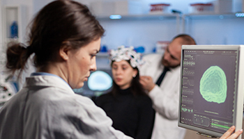
To address this, the hospital integrated our AI-powered Brain Tumor Segmentation Platform, which leverages deep learning models trained on thousands of MRI datasets. The system automatically identifies and highlights tumor regions in high-resolution scans.

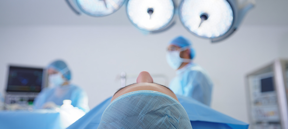
The AI solution streamlined the pre-surgical process by reducing planning time by 40%, enabling clinicians to focus on critical surgical decisions rather than manual data interpretation. With real-time visualization and integration into existing PACS systems, the hospital achieved improved accuracy, reduced workload, and enhanced patient safety in neurosurgical planning.

Overview Image
Challenges

With complex MRI data and rising surgical demands, Mivora Neuro Hospital faced:

  • Long planning times due to manual segmentation
  • Variations in tumor boundary identification
  • Risk of incomplete visualization of tumor regions
  • High workload on radiologists
  • Delays in surgical decision-making
No items found.
Solutions

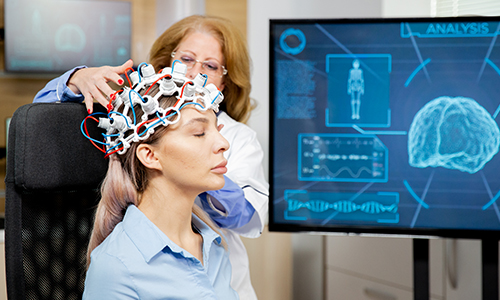
The AI-powered system was developed to assist neurosurgeons by delivering precise, automated brain tumor segmentation from MRI scans.

  • Automated Tumor Segmentation
    Instantly analyzes MRI scans to delineate tumor regions, reducing manual annotation time.
  • 3D Visualization & Mapping
    Provides surgeons with high-resolution 3D reconstructions of tumor structures and surrounding tissues.
  • Integration with Surgical Tools
    Seamlessly integrates with neuronavigation systems to support intraoperative decision-making.
  • Confidence Mapping
    Displays confidence scores for each segmented area, allowing clinicians to validate and adjust surgical approaches.
No items found.
Outcome
  • Reduced pre-surgical planning time by 60%
  • Improved accuracy in tumor boundary detection by 25%
  • Enhanced visualization leading to safer, more precise surgeries
  • Empowered neurosurgeons with real-time, data-driven surgical insights
Results

Delivered precise, data-driven insights that improved surgical accuracy, reduced planning time, and enhanced patient outcomes.

95% segmentation

Tumor Boundary Precision

40% faster

Surgical Planning Time

2x improvement

Surgeon Decision Efficiency
Avatar Image

Dr. Amanda Liu

Diagnostic Imaging Lead

AI gives us an extra set of expert eyes. It spots subtle indicators in scans that even trained specialists might miss.

Other case studies

Your form has been submitted successfully. Thank you!
Please double-check your information and try again. If the issue continues, you can also email us directly at support@yourdomain.com

Transform Healthcare with the power of AI

Join the future of patient-centered care. Connect with our team to discover how our AI innovations can revolutionize your healthcare operations.

Avatar ImageAvatar ImageAvatar ImageAvatar Image
Icon
Rated 4.5/5 from 400+ reviews
More Templates